Assignment #2 - Scientific Paper Review
Phuc Van Pham & Loan Thi-Tung Dang &
Uyen Thanh Dinh & Huyen Thi-Thu Truong &
Ba Ngoc Huynh & Dong Van Le & Ngoc Kim Phan
2013
2013
Introduction
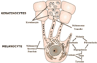
When skin ages due to UV radiation, it is referred to as photoaging. Photoaging occurs when fibroblasts and keratinocytes proliferation is inhibited, also collagen and fibronectin expression is inhibited. It also activates collagenases, which breakdown collagen, such as matrix metalloproteinase-1. All of these factors cause wrinkles, the major characteristic of skin aging. Pham et al (2013) used intracellular umbilical cord extracts, extracellular umbilical cord extracts and umbilical cord-derived stem cell extracts to test the effects on fibroblasts, keratinocytes and melanocytes. Fibroblasts are responsible for producing collagen in the dermis of the skin. Keratinocytes make up 90% of the skins epidermis which is used for protection from pathogens, heat, UV radiation and water loss. Melanocytes are located in the bottom layer of the epidermis which produce melanin. Melanin affects the pigment of your skin and absorbs UV light and prevents exposure to the dermis layer.
Figure 1: Skin aging
Figure 2: Young skin versus older skin
Figure 3: Melanocytes and Keratinocytes Figure 4: Fibroblasts in dermis
Results
Pham et al (2013) had very positive results with their experiment. They collected umbilical cords under consent and isolated the three skin cell types. Many experiments have been done with placental extracts on skin aging, but few from umbilical cord extracts. There are at least four areas of the umbilical cord that are stem-cell rich. These include the cord lining membrane, Wharton's jelly, umbilical cord vein and umbilical cord blood. Pham et al (2013) prepared 25 formulae, five being the control. They found that 7.5% and 10% of formula 35 stimulated the proliferation of fibroblasts significantly more than the control. Therefore, they chose the 7.5% for the optimal concentration to continue with the experiment. Formula 35 contains both extracellular and intracellular umbilical cord extracts. In the keratinocytes, 7.5% of formula 35 also increased the proliferation significantly more than the control. Next, Pham et al (2013) tested formula 35 on collagen 1, fibronectin and MMP-1. They found that this formula strongly inhibited MMP-1, while significantly stimulating collagen 1. Formula 35 did not show a change in fibronectin or on the expression of tyrosinase, which is important in the synthesis of melanin. Overall, they proved that by stimulating keratinocyte and fibroblasts proliferation, they can inhibit MMP-1 and stimulate collagen 1, reducing skin aging.
Figure 5: The 25 formulae used by Pham et al (2013)
Critique


